Publications by application
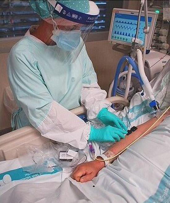
Clinical and Rehabilitation
The medical world is continuously searching for new ways to help people in need. Over the years, the NIRS devices from Artinis have shown to be well suited for research in a clinical setting.
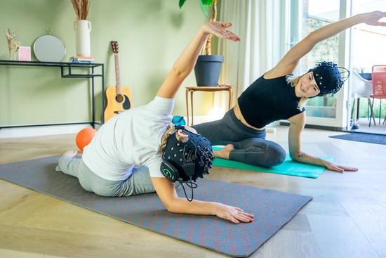
Hyperscanning
Hyperscanning is defined as simultaneously measuring the brain activity of multiple subjects. Social interactions are a crucial part of human life. Understanding the neural underpinnings of social interactions is a challenging task that the hyperscanning method has been trying to tackle over the last two decades.

Hypoxia and Altitude studies
A constant supply of oxygen to our muscles and organs is essential for the maintenance of proper body functioning. However, we frequently encounter conditions where the oxygen demand by tissue exceeds the supply, thereby leading to a state of oxygen deprivation, also called hypoxia.
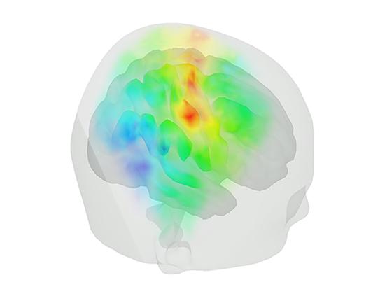
Motor cortex
fNIRS stands for functional near infrared spectroscopy. The functional component comes from the fact that our fNIRS devices are capable of assessing brain activity. This is done by measuring changes in oxyhemoglobin and deoxyhemoglobin, which reflect local brain activity.
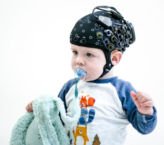
Neonatal
The complex architecture of the human brain develops in an ongoing process that starts in the womb and continues into adulthood. In the first few years of human life, more than 1 million new neural connections are formed every second.
Neurostimulation fNIRS
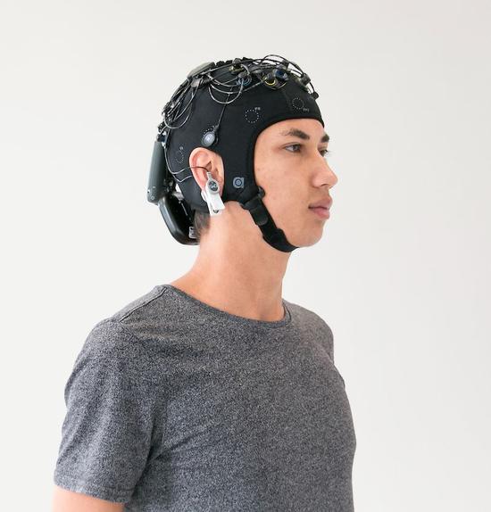
As fNIRS measures brain activity based on light, no interference occurs when combined with neurostimulation methods that use magnetic (TMS) or direct current stimulation (tDCS). Moreover, this makes fNIRS an ideal brain imaging modality in research where the brain has to be stimulated and local activity simultaneously quantified.
NIRS-EEG
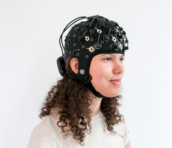
There are multiple brain imaging modalities available nowadays. But how do you decide which modality fits your research best? fNIRS is known for the fact that it is non-invasive, portable, and has a good spatial resolution.
Parietal cortex
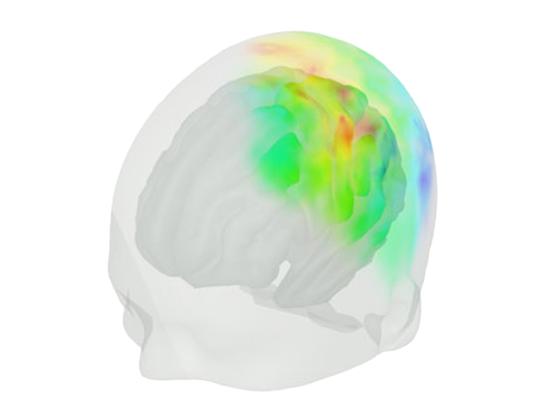
fNIRS stands for functional near infrared spectroscopy. The functional component comes from the fact that our fNIRS devices are capable of assessing brain activity. This is done by measuring changes in oxyhemoglobin and deoxyhemoglobin, which reflect local brain activity.
Prefrontal cortex
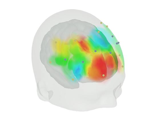
fNIRS stands for functional near infrared spectroscopy. The functional component comes from the fact that our fNIRS devices are capable of assessing brain activity. This is done by measuring changes in oxyhemoglobin and deoxyhemoglobin, which reflect local brain activity.

Sports science
The field of sport Science is one of the first research fields where NIRS found wide use application. In sport Science it is critical to have insight in the local muscle oxygen consumption.
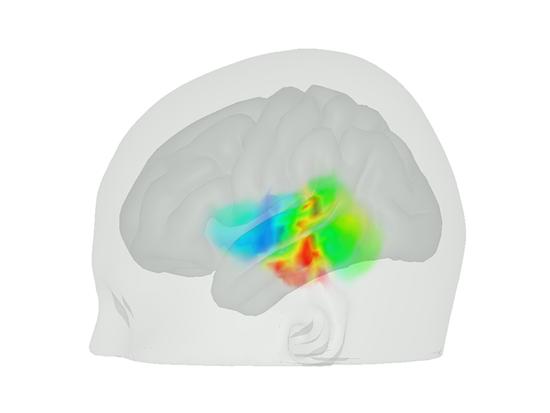
Temporal cortex
fNIRS stands for functional near infrared spectroscopy. The functional component comes from the fact that our fNIRS devices are capable of assessing brain activity. This is done by measuring changes in oxyhemoglobin and deoxyhemoglobin, which reflect local brain activity.
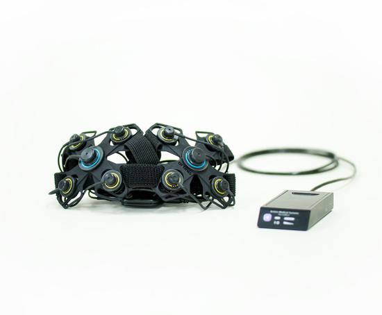
Urology
Over the years NIRS has been utilized in many different fields of research. Recently, NIRS has also found use in the field of urology. Indications show that bladder blood flow and oxygenation differ in health and disease.
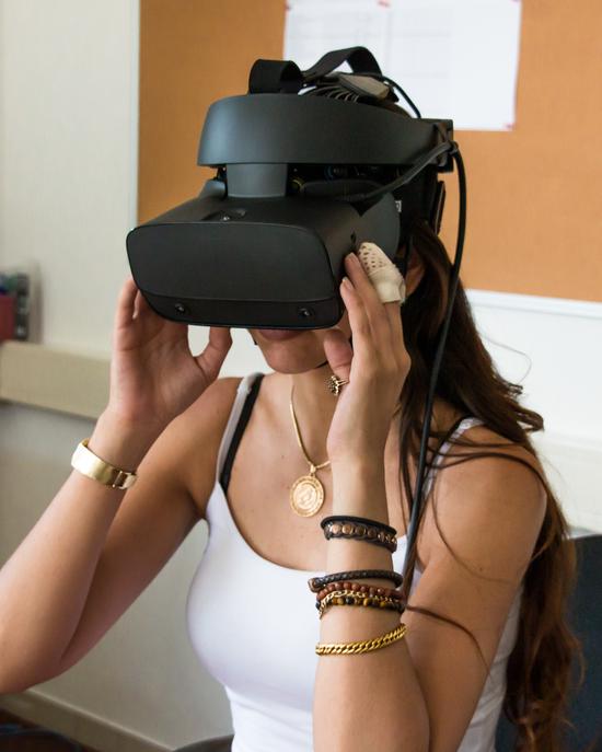
Virtual Reality
VR is a simulated experience that can be sometimes almost indistinguishable from real-world experiences. Since the fNIRS devices produced by Artinis can be easily combined with VR headsets, researchers have utilized this by assessing brain activity while their subjects are immersed in a VR world.
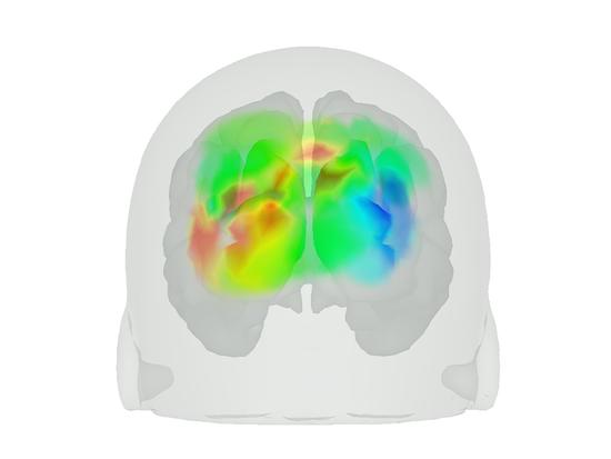
Visual cortex
fNIRS stands for functional near infrared spectroscopy. The functional component comes from the fact that our fNIRS devices are capable of assessing brain activity. This is done by measuring changes in oxyhemoglobin and deoxyhemoglobin, which reflect local brain activity.
Brain Computer Interfaces
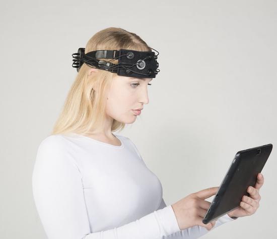
BCI devices are gaining more and more popularity because of the exciting potential they hold. The non-invasive nature of our fNIRS systems in combination with being easy to set up, makes them a great brain imaging modality for use in BCI applications.
Featured Publications
Cerebral oxygenation responses to head movement measured with near-infrared spectroscopy
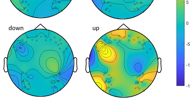
Motion is disruptive to neuroimaging methods. Motion artefacts range from large amplitude and short frequency spikes to drifts in amplitude causing cofounds in the analysis or completely invalidating any analysis, leading to epoch exclusion of data. However, we can only acquire ecologically robust information if subjects are engaging in natural interaction with their environment. Even more so in the case of sports, infants or motor disorder afflicted populations, where movement will happen. We proposed to study the relationship between channel location and Inertial Measuring Unit (IMU) quantified head movements, in order to better understand their effects in fNIRS data. Cerebral oxygenated (O2Hb) and deoxygenated (HHb) haemoglobin were measured bilaterally in prefrontal to frontal and occipital to temporo-parietal regions of healthy individuals. All participants performed controlled head movements in four conditions: Up, Down, measured by pitch IMU values; and Left, Right, measured by yaw IMU values, in varying degrees of movement. We analysed amplitude and coefficient of determination of O2Hb and HHb, within conditions and channel coordinates across subjects. Our results show that smaller angle magnitude movements (bellow 60 degrees in rotation) are significantly different than larger angle magnitude movements (above 75 degrees in rotation) with a p value of 0.0073; and that the Up condition is significantly different than other movement directions with a p value of 0.0001. We conclude that movement artefacts do not depend on area of measurement for the movement conditions studied. We recommend the application of threshold values for the future with the use of the IMU, by ignoring the effects of lower magnitudes of movement, while correcting or removing larger magnitudes. In future motion artefact removal, we recommend using an IMU for optimal head motion correction of cerebral oxygenation signals


